Structure
Calmodulin (CaM) is a small, calcium-binding protein consisting of a single peptide chain, 148 amino acid residues (52 of which are charged) and a molecular weight of 16,700. CaM consists of three domains, i.e., two calcium binding domains connected by a central helix. Domain 1 consists of residues 1-64, domain 2 has residues 65-92 and domain 3 has residues 93-148. Domains 1 and 3 are identical in packing density and but the packing density of the central helix/domain 2 is much higher due to extensive hydrogen bonding.
CaM is largely composed of alpha helixes (63%) and upon calcium binding has a "dumbbell" shape owing to its two globular lobes that are connected via a long and exposed alpha helix. Each of the lobes contains two Ca2+ ion binding sites so that there is a total number of four Ca2+ binding sites per calmodulin molecule. The Ca2+ binding sites are helix-loop-helix domains (EF hands) that are similar to those of other calcium binding proteins like troponin-C. The interconnecting alpha helix is thought to be responsible for interactions between calmodulin and other proteins, drugs etc., and may also play a role in Ca2+ binding cooperativity of the molecule.

Figure 1. The basic 3-dimensional structure of Calmodulin without the bound calcium atoms. This form of CaM is inactive. The alpha-helixes are shown in red and the beta sheets are shown in blue. The white regions are the random coils or the nonrepetitive secondary structures. The overall length of the molecule is ~65A0 while the EF hands have approximate dimensions of 25x20x20 A0.
The calmodulin molecule consists of seven alpha helices that consist of the residues as shown below.
Table 1. The Alpha-Helixes of CaM and their Respective Amino Acid Residues
|
Alpha-Helix
|
Amino Acid Residues
|
|
I
|
7-19
|
|
II
|
29-39
|
|
III
|
46-55
|
|
IV
|
65-92
|
|
V
|
102-112
|
|
VI
|
119-128
|
|
VII
|
138-148
|
This info. is courtesy of Babu et al., 1985.
The seven alpha-helices and their relative positions to one another are shown in Figures 2 and 3.
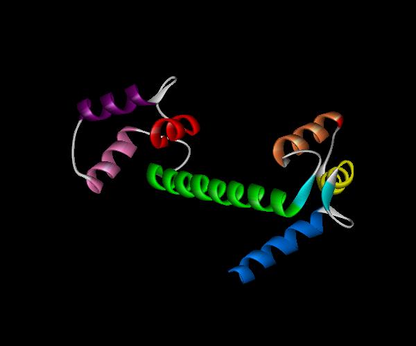
Figure 2. CaM with all the alpha helices shown in different colors. Helix I=dark blue, Helix II= yellow, Helix III=orange, Helix IV=green, Helix V=pink, Helix VI=purple, Helix VII=red.
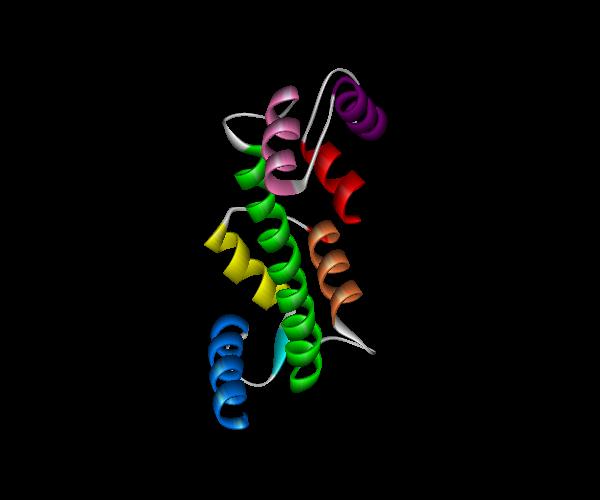
Figure 3. Yet another view of calmodulin's seven alpha helices. Helix I=dark blue, Helix II= yellow, Helix III=orange, Helix IV=green, Helix V=pink, Helix VI=purple, Helix VII=red.
CaM is largely stabilized by many interactions among the helices. There is also hydrogen bonding between the calcium binding sites in each dumbbell. In the first half of the molecule amino acid residues 25-29 run antiparallel to residues 61-65 and the resulting beta sheet is kept intact by hydrogen bonding between Gly 25 and Asp 64 and between Ile 27 and Thr 62. In the second half of the molecule residues 99-101 and 135-137 run antiparallel to each other and the beta sheets is held together by hydrogen bonding between Ile 100 and Val 136. Similar arrangements are also found in other calcium binding proteins like parvalbumin and intestinal calcium binding protein.
CaM also has four Ca2+ binding sites (helix-loop-helix domains/EF hands) that each consist of twelve amino acid residues: 20-31, 56-67, 93-104 and 129-140 (Babu et al., 1985). The first nine amino acid residues of each domain form the first helix and loop of the domain and the remaining three residues mark the beginning of the second helix. In each binding site the Ca2+ ions are displaced from the center of the loop towards the amino terminal domain and the second helix helps to orient the last three residues so that they can all participate in coordinating Ca2+ binding.
Figure 4 shows the Ca2+ binding domains of calmodulin.

Figure 4. The Ca2+ binding domains of calmodulin are shown in green in the above graphic. Notice that there are two Ca2+ binding sites for each "dumbbell" of the molecule, which are connected via the central helix, i.e., Helix IV, shown in yellow.
Calcium binding domains I (residues 20-31) and IV (residues 129-140) have the helix-loop-helix conformations that are similar to other calcium binding proteins like parvalbumin and intestinal calcium binding protein. Domains II (residues 56-67) and III (residues 93-104) of CaM, although they have the same helix-loop-helix conformations are different in that they are connected via Helix IV which it the longest alpha helix of the molecule shown in yellow above). Helix IV provides the carboxy terminal for domain II and the amino terminal for domain III.
Shown below is a fully saturated calmodulin molecule with all four of its Ca2+ binding sites occupied by the metal ions.

Figure 5. A fully saturated calmodulin molecule with Ca2+ ions shown as green spheres. Notice how in accordance with the two binding sites per dumbbell there are two metal ions bound per dumbbell. In its fully saturated form, calmodulin can activate and interact with its target enzymes. The two calcium binding sites in each dumbbell are related to each other by an approximate two-fold axis and are ~11.3 A0 apart (Babu et al., 1985).